Contributions
- We developed the fastest, at the time, 3D MRI data acquisition technique for obtaining whole-brain datasets in less than 0.9secs on clinical MRI scanners, for the purpose of event-related fMRI with sub-second temporal resolution (2003). We achieved that by combining principles of echo-shifting and parallel imaging.
- We have published some of the early articles demonstrating the principles of resting state fcMRI, and showed that functional connectivity is characterized mainly by frequencies lower than 0.1 Hz.
- We showed for the first time that synchronous low frequency fluctuations observed during resting state are also present when other brain regions are activated (during task-related fMRI) (2000).
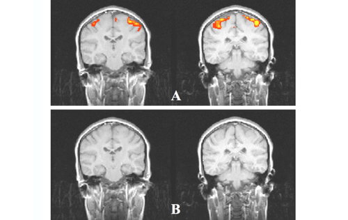
- We have demonstrated the use of independent component analysis (ICA) to remove activation from data acquired during the performance of a task in order to produce better-controlled "resting" state conditions.
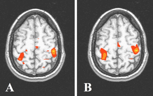
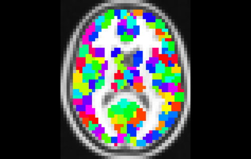
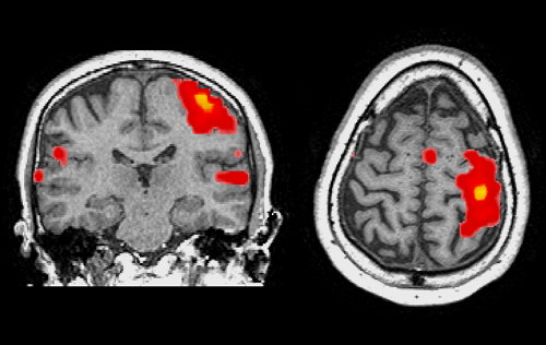
Functional MRI (fMRI) provides information about the location and temporal characteristics of neuronal activity. This is achieved by collecting a time series of brain MR image volumes while the subject is presented a stimulus or performs a task, and investigating local signal changes following neuronal activity. Activated brain regions are identified as those with a signal time-course that fits well the expected hemodynamic response. In functional connectivity MRI (fcMRI), the subject is typically at rest (no stimulus), and functionally connected brain regions are identified as those that exhibit synchronous low frequency fluctuations.
Selected Related Publications
- Arfanakis K, Cordes D, Haughton VM, Moritz CH, Quigley MA, Meyerand ME. Combining independent component analysis and correlation analysis to probe interregional connectivity in fMRI task activation datasets. Magn Reson Imaging. 2000;18:921-930.
- Cordes D, Haughton VM, Arfanakis K, Wendt GJ, Turski PA, Moritz CH, Quigley MA, Meyerand ME. Mapping functionally related regions of brain with functional connectivity MR imaging. AJNR Am J Neuroradiol 2000;21:1636-1644.
- Cordes D, Haughton VM, Arfanakis K, Carew JD, Turski PA, Moritz CH, Quigley MA, Meyerand ME. Frequencies contributing to functional connectivity in the cerebral cortex in "resting-state" data. AJNR Am J Neuroradiol 2001;22:1326-1333.
- Han D, Buchman AS, Arfanakis K, Fleischman DA, Bennett DA. Functional connectivity networks associated with chronic musculoskeletal pain in old age. Int J Geriatr Psychiatry 2013;28:858-867.
- Han SD, Boyle PA, Arfanakis K, Fleischman DA, Yu L, Edmonds EC, Bennett DA. Neural intrinsic connectivity networks associated with risk aversion in old age. Behav Brain Res 2012;227:233-240.